MetalloFluor™ Series

FerroFarRed™

[Labile ferrous ion detecting probe]

650-750 nm:Far red

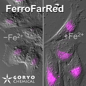
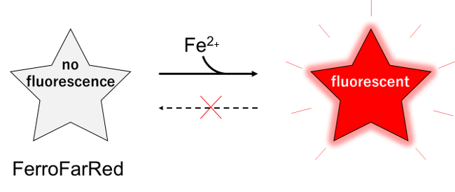
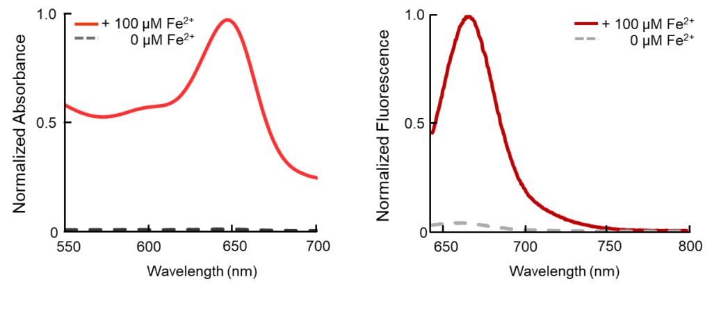
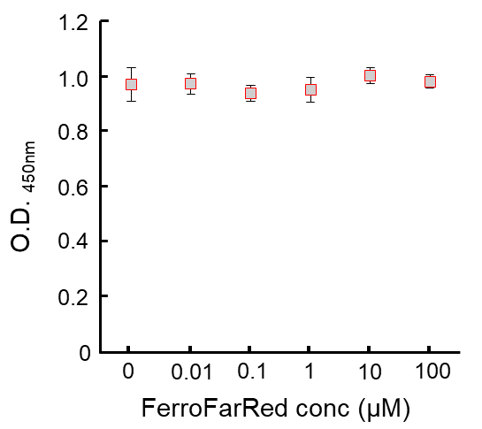
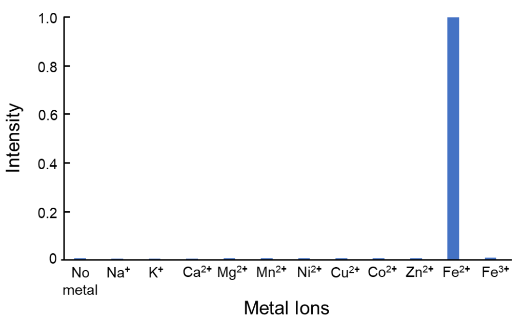
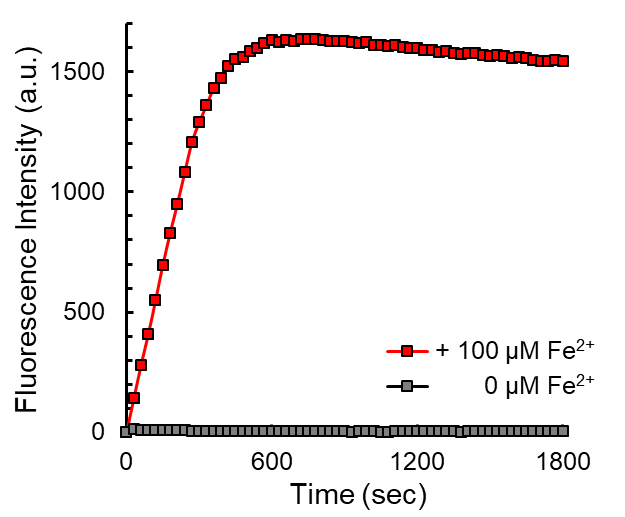
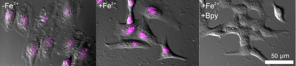
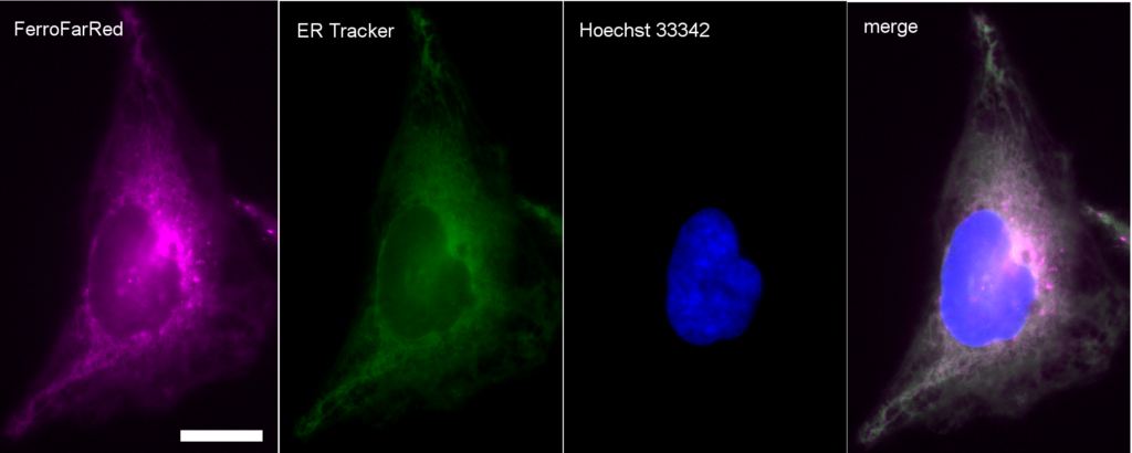
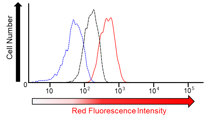
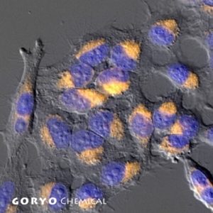
FerroFarRed (also known as SiRhoNox-1 or ER-SiRhoNox) is a fluorescent probe that specifically detects labile iron (II) ions (Fe2+). This probe is designed to selectively react with only Fe2+ separately from other metal ions and irreversibly turns into a far-red fluorescent substance. It mainly localizes inside endoplasmic reticulum (ER). It can also be used with a flow cytometer equipped with a red laser.

This probe does not have chelating effect.

Available through Merck KGaA (Darmstadt, Germany) as:
SCT037  BioTracker™ Far-red Labile Fe2⁺ Dye 


Products

Code No. Product Name Size Merck CAT No. Merck ( Millipore / Sigma Aldrich )
Product Name
GC903-01 FerroFarRed™ 50 nmol × 5 SCT037 BioTracker Far-red Labile Fe2⁺ Dye 

Downloads

  • Protocol

  • SDS

  • Product Information

    Print

    Propertities of FerroFarRed

    Product Name Target Cell permeability Reactivity Absmax (nm) FLmax (nm)
    FerroFarRed Labile Iron (Fe2+) Yes Irreversible 646 662

    FerroFarRed is a fluorescent probe that specifically detects labile iron (II) ions (Fe2+). Fluorescence intensity does not increase with iron (III) ions (Fe3+) or other divalent metal ions.

    Principle of reaction

    FerroFarRed is a blue substance with no fluorescence. FerroFarRed react with Fe2+ and irreversibly turns into a far-red fluorescent substance. (Absmax = 646 nm, FLmax = 662 nm)


    Absorption and fluorescence spectrum


    Absorption (left) and fluorescence (right) spectra of FerroFarRed.
    Reaction with Fe2+ significantly increases the fluorescence intensity.

    Cytotoxicity

    FerroFarRed shows almost no cytotoxicity at concentrations up to 100 μM (~20 times that of generally used concentrations).


    Metabolic activity of HeLa cells in various concentrations of FerroFarRed measured by the CCK-8 assay (n = 3; ±S. D.).

    FerroFarRed was diluted in DMSO at various concentrations and then each solution was added in the medium (containing 0.1% DMSO as a cosolvent).
    After 3 hours, cells were washed twice with HBSS, then fresh medium was replaced. Metabolic activity of HeLa cells was measured by CCK-8 assay at 24 hours after washing (n=3; ±S. D.).


  • Reactivity of FarroFarRed

    Print

    Reactivity of FerroFarRed

    Metal Ions Reaction Specificity

    A pronounced increase in fluorescence occurs only in the presence of Fe2+.

    Reactivity of FerroFarRed with various metal ions. Relative fluorescence intensity to that upon a reaction with Fe2+.

    Measurement conditions
    Fluorescence of 5 μM FerroFarRed in HEPES buffer (0.05 M, pH 7.4, containing 0.1% DMSO as a cosolvent) was measured after reaction with various metal ions for 60 minutes at 37°C. (The concentration of Na+, K+, Ca2+ and Mg2+ is 1 mM and that of the other metal ions is 20 μM)
    The fluorescence intensities measured at 665 nm, with excitation at 630 nm by using a microplate reader (TECAN infinite M200Pro).


    Reaction Rate

    Changes in fluorescence intensity of 5 μM FerroFarRed with or without 100 µM of Fe2+.

    Fluorescence intensity of 5 μM FerroFarRed in HEPES buffer (0.05 M, pH 7.4, containing 0.1% DMSO as a cosolvent) was recorded every 30 seconds with a microplate reader (TECAN infinite M200Pro). The fluorescence intensities measured at 665 nm, with excitation at 630 nm.


  • Imaging and measurement examples using FerroFarRed

    Print

    Imaging and measurement examples using FerroFarRed

    A cell imaging example by fluorescence microscopy

    Fluorescence signal was increased in cells treated with Fe2+ (middle) compared with control cells (left). The Fluorescence signal was decreased by adding Bpy (2,2′-Bipyridyl), Fe2+ chelator, in addition to Fe2+ (right).

    Live cell imaging of Fe2+ in HeLa cells. The respective panel shows control cells (left), cells treated with Fe2+ (middle) and cells co-treated with Fe2+ and Bpy (right). HeLa cells cultured for 30 minutes in serum-free medium containing 100 μM Fe(SO4)2(NH4)2 and HeLa cells without loading Fe2+. The cells were rinsed with HBSS to remove extracellular Fe2+ions before reacting with 5 µM FerroFarRed at 37oC for 1 hour. 1 mM Bpy was co-treated with 5 μM FerroFarRed. The cells were observed under a fluorescence microscope using a 590-650 nm excitation filter and a 660-740 nm fluorescence filter. The magenta pseudo color shows the fluorescent image of FerroFarRed. Magenta pseudo color was overlaid to grayscale images of DIC.


    Subcellular localization of FerroFarRed

    HeLa cells stained with ER Tracker (green) and Hoechist 33342 (blue) was reacted with FerroFarRed (magenta). FerroFarRed mainly localizes in endoplasmic reticurrum.


    A measurement example by flow cytometry

    FerroFarRed is also applicable to flow cytometry using a red laser.

    Flow cytometric analysis on HepG2 cells. The respective histograms show control cells (black dashed line), cells treated with Fe2+ (red solid line) and cells co-treated with Fe2+ and Bpy (blue dotted line). For loading Fe2+, HepG2 cells cultured for 30 minutes in serum-free medium with 100 μM Fe(SO4)2(NH4)2. After the cells rinsed with HBSS to remove extracellular Fe2+, HepG2 cells reacted with 5 µM FerroFarRed at 37oC for 1 hour. 1 mM Bpy was co-treated with 5 μM FerroFarRed. Cells were washed with PBS and analyzed using FACSVerse™ (Becton Dickinson). FerroFarRed was excited using a red laser (640 nm) and measured using an allophycocyanin (APC) fluorescence filter.

FAQ

  • Q Does the probe only detects Fe2+ in Golgi/ER?
    A

    The probes are known to be localized either in Golgi or in ER, however,  it has been considered that they also detect Fe2+ in the cytoplasmic pool. Please note that the definitive evaluation for this point has not been reported, yet. 

  • Q Can FerroFarRed, FerroOrange be used in fixed samples?
    A

    These reagents could not be used for paraffin slices.

    These reagents could only be used in mild fixation conditions such as 3% paraformaldehyde (PFA) in PBS for 10 min at 4°C.
    The fluorescence intensity of the samples which were fixed for 20 minutes or more, or fixed at room temperature, was significantly decreased compared with unfixed samples.

    Please note that it might be difficult to react these probes with fixed samples. Apply the reagent first, then try fixation.

  • Q Which of the ferrous detecting reagents is the most appropriate for my purpose?
    A

    FerroOrange is the most sensitive probe for ferrous ions. In contrast, FerroFarRed is suitable for flow cytometry with red laser.
    Regarding the number of the publications, FeRhoNox-1 (RhoNox-1) might be the best to reproduce experiments.

    Code Number Product Name Exmax (nm) Emmax (nm) Microscopy Flow cytometry
    (blue laser)
    Flow cytometry
    (red laser)
    Plate reader
    GC901 FeRhoNox-1 540 575
    GC903 FerroFarRed 646 662
    GC904 FerroOrange 542 572

    We recommend to use a green laser (ex. 532 nm) to excite FeRhoNox-1 or FerroOrange.

  • Q My question is not in this FAQ list.....

Reference

H.Jiang , R. K. Muir, R. L. Gonciarz, A. B. Olshen, I. Yeh, B. C. Hann, N. Zhao, Y. H. Wang , S. C. Behr, J. E. Korkola, M. J. Evans, E. A. Collisson, A. R. Renslo (2022)
J. Exp. Med. 219: e20210739. DOI: 10.1084/jem.20210739

D. Y. Kang, N. Sp, E. S. Jo, J. M. Lee. K. J. Jang (2021)
Cells 10: 2549-2562 DOI: 10.3390/cells10102549

Fumoto, E. Kinoshita,K. Ohta, K. Nakamura,T. Hirayama, H. Nagasawa, D. Hu, K. Okami, R. Kato, S. Shimokawa, N. Ohira, K. Nishimura, H. Miyamoto, T. Tanaka, S. Kawakami, K. Nishida (2020)
Pharmaceutics 12: 1070 DOI: 10.3390/pharmaceutics12111070

Z.Li, L. Jiang, S. H. Chew, T. Hirayama, Y. Sekido, S. Toyokuni (2019)
Redox Biol. 26: 101297 DOI: 10.1016/j.redox.2019.101297

D. Cui, M. Arima, T. Hirayama, E. Ikeda (2019)
Exp. Cell Res. 379 : 166-171 DOI: 10.1016/j.yexcr.2019.04.003.

K. Sato, L. Shi, F. Ito, Y. Ohara, Y. Motooka, H. Tanaka, M. Mizuno, M. Hori, T. Hirayama, H. Hibi, S. Toyokuni (2019)
J. Clin. Biochem. Nutr. 65 : 8-15 DOI : 10.3164/jcbn.18-91

T. Imai, S. Iwata, T. Hirayama, H. Nagasawa, S. Nakamura, M. Shimazawa  H. Hara (2019)
Sci. Rep. 9: 6228-6243 DOI: 10.1038/s41598-019-42370-z

T. Hirayama, A. Miki, H. Nagasawa (2018)
Metallomics. in press DOI: 10.1039/c8mt00212f

K. Sakamoto, T. Suzuki, K. Takahashi, T. Koguchi, T. Hirayama, A. Mori, T. Nakahara, H. Nagasawa, K. Ishii (2018)
Exp. Eye Res. 171: 30-36 DOI: 10.1016/j.exer.2018.03.008

T. Hirayama, H. Tsuboi, M. Niwa, A. Miki, S. Kadota, Y. Ikeshita, K. Okuda, H. Nagasawa (2017)
Chem. Sci.
8: 4858-4866 DOI: 10.1039/c6sc05457a