GlycoFluor™ Series

GlycoYELLOW™ –βGal

[β-galactosidase activity detecting probe]

540-570 nm:Yellow

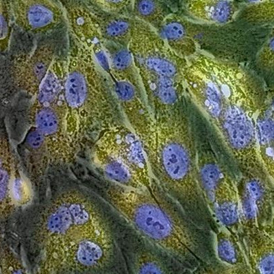
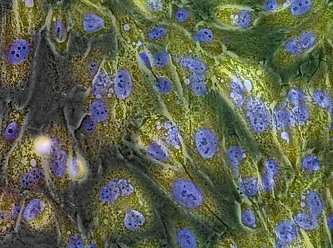
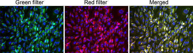
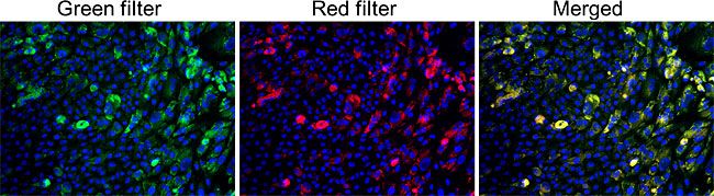
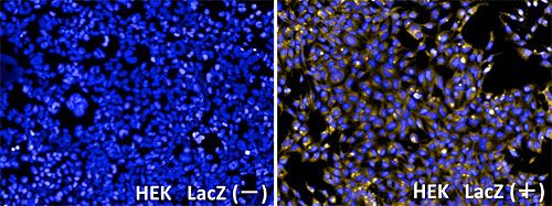
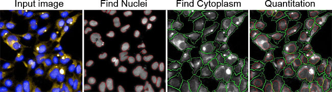
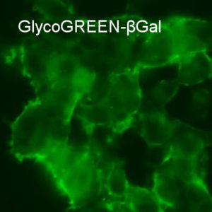
GlycoYELLOW-βGal is a fluorescent ligand for β-galactosidase. It shows almost no fluorescence but fluoresces upon the reaction with β-galactosidase. It can be applied to fluorescence imaging and selection of lacZ-gene expressing cells and tissues. In addition, it can be used to detect senescence-associated β-galactosidase (SA-β-Gal) activities.


Available through Merck KGaA (Darmstadt, Germany ) as:
SCT024 BioTracker™ 543 Yellow β-Gal Dye


Products

Code No. Product Name Size Merck CAT No. Merck ( Millipore / Sigma Aldrich )
Product Name
GC601 GlycoYELLOW™ –βGal 50 μg × 10 SCT024 BioTracker 543 Yellow β-Gal Dye

Downloads

  • Protocol

  • SDS

  • Product Information

    Print

    About GlycoYELLOW-βGal

    Upon the reaction with β-galactosidase, it shows yellow fluorescence.

    Properties of GlycoYELLOW-βGal

    Product Name target reaction Absmax (nm) FLmax (nm)
    GlycoYELLOW-βGal β-galactosidase activity irreversible 525 543


    Spectra

    Fluorescence spectra before and after the reaction with β-galactosidase. Reaction was at 37oC for 30 minutes.

  • Imaging radiation-induced senescence with GlycoYELLOW-βGal

    Print

    Imaging radiation-induced senescence with GlycoYELLOW-βGal

    Data were kindly provided by Keiji SUZUKI, Ph.D, Department of Radiation Medical Sciences Atomic Bomb Disease Institute, Nagasaki University.

    Live cell imaging of radiation-induced senescent cells using GlycoYELLOW-βGal. Yellow signal indicates β-galactosidase (β-Gal) activity in living cells.


    Live cell imaging movie showing β-Gal activity in thyroidal gland follicular cells 10 days after the irradiation of 10 Gy of γ-ray. Cell cycle was also monitored with Fucci system.
    G1 arrest caused by radiation-induced senescence is visualized by the red-color of the nucleus. Red signal increase in the cytoplasm indicates SA-β-gal, which is monitored with GlycoYELLOW-βGal. Observation period of 24-hours were recorded.

    Staining of living thyroidal gland follicular cells

    Human thyroidal gland follicular cells were irradiated with 10 Gy of γ-ray, cultured for 10 days and reacted with GlycoYELLOW-βGal for 30 minutes. SA-β-gal was detected as yellow fluorescence (both in red and green channel). In this condition, ~100% cells were also positive in X-gal staining.

    On the other hand, in the area where cells were still dividing (bottom-left corner), β-gal activity (yellow signal) was weak.  This result indicates that GlycoYELLOW-βGal can detect radiation-induced cellular senescence (SA-β-gal).


  • Screening β-galactosidase gene marker by using GlycoYELLOW-βGal

    Print

    Screening β-galactosidase gene marker by using GlycoYELLOW-βGal

    Introduction

    β-galactosidase (β-gal) activity has been widely used as a senescent marker (SA-β-Gal) or reporter gene (lacZ). Conventional X-Gal staining, analysis of β-Gal activity at single-cell level is difficult due to poor sensitivity.  GlycoYELLOW-βGal, a highly sensitive fluorescent probe for β-Gal activity enables single-cell level detection with high S/N ratio. This probe is applicable for both live and fixed cells. Therefore, it is suitable for pharmacological tests, including cell imaging-based screening system as well as detection of cellular senescence. To test the possibility, we analyzed β-gal activity of LacZ-transfected cells at single-cell level using Operetta™ (PerkinElmer , USA).


    Fixation and reaction of HEK293 cells (either LacZ-gene transfected, ie. LacZ+, or not, ie. LacZ-)

    1. Added 50 µL poly-L-lysine to each well of 96 well plate and incubated at 37℃ overnight in a 5% CO2 incubator.
    2. Removed poly-L-lysine and washed 96 well plate with HBSS twice.
    3. Tripsinized and dissociated HEK293 cells were filterated through a cell strainer (40 μm nylon mesh). Then the cells were seeded to 96 well plate (5×10⁴ cell/ml, 200 µl/well) and incubated for 37℃ overnight in a 5% CO2 incubator.
    4. Diluted GlycoYELLOW-βGal 1 vial (50 µg) in 93.3 µL DMSO to prepare 1 mM stock solution.
    5. Diluted the 1 mM stock solution with HBSS to 5 µM (reaction solution). Also added 5000 times diluted Hoechst33342 (1 mg/ml) for nuclear staining to the reaction solution.
    6. Removed the culture medium in the wells and gently rinsed cells once with HBSS.
    7. Added 200 µl of the reaction solution to each well and incubated at 37°C for 60 min.
    8. Removed the culture medium on the dish and gently rinsed the cells twice with the observation buffer.
    9. Fixed the cells for 10 to 30 minutes with 3% of paraformaldehyde.
    10. After the fixing, gently rinsed the cells once with HBSS.
    11. Added fresh HBSS and observation with Operetta™ (PerkinElmer). 10× long WD lens, ex. 360-400 nm/em. 410-480 nm for Hoechst 33342, ex. 520-550 nm/em. 560-630 nm for GlycoYELLOW-βGal were used.


    Image analysis of GlycoYELLOW-βGal reacted cells

    Following psudocolor images were overlayed; blue: Hoechist 33342 and yellow: GlycoYELLOW-βGal.
    The fluorescence intensity of GlycoYELLOW-βGal was much higher in the LacZ expressing cells (HEK LacZ (+)) than that in the non-expressing cells (HEK LacZ (-)).

    The result of imaging analysis via Harmony software on Operetta™
    (Left) Comparing averaged fluorescence intensity of LacZ(+/-) cells. HEK LacZ (+) cells showed more than 7.5 times higher fluorescence intensity compare to that of HEK LacZ (-) cells. (Right) The number of analyzed cells.  Number of cells are obtained by counting the number of nuclei. The number of cells are sufficient for the statistical analysis.

    Details of cell analysis

    The Images were acquired using Operetta™ High Contents Imaging System (PerkinElmer, Hopkinton, MA) with 10x objective with appropriate optical filters. The obtained images were analyzed with Harmony™ software (PerkinElmer). In brief, the individual cells in each field of view (FOV) were recognized by using prebuild software functions as follows: DAPI stained nuclei were recognized by “Find Nuclei” function, and then cytoplasmic region were subsequently detected by “Find Cytoplasm” function. The fluorescence intensity derived from GlycoYELLOWTM-βGal in cytoplasm region was quantitated in every cell in FOV. Each step of the image analysis is summarized in Figure 3.

    The analysis  with Harmony™

    Input image: Representing image of GlycoYELLOW-βGal stained cells acquired on Operetta™.
    Find Nuclei: Nuclei recognized in input image with “Find Nuclei” function.
    Find Cytoplasm: Cytoplasm regions recognized in input image with “Find Cytoplasm” function.
    Quantitation: GlycoYELLOW-βGal positive signal detected in the FOV.


    For further information or questions, please contact Goryo Chemical, Inc.


FAQ

  • Q Give me a selection guide of the β-galactosidase probes.
    A

    We have three β-galactosidase probes, GlycoGREEN-βGal, GlycoGREEN-βGal, and TokyoGreen-βGal . For cell imaging, we recommend GlycoGREEN-βGal, for plate-reader assays or for enzyme kinetics measurements, we recommend TokyoGreen-βGal as a first choice.

    Sensitivity of the probe isGlycoGREEN-βGal ~  TokyoGreen-βGal > GlycoYELLOW-βGal. 
    For cell imaging, we recommend GlycoGREEN-βGal because its cell retention is relatively better, however, generated fluorophore also leaks from the cells and can be incorporated into other cells, users should carefully check the result when they observe heterogeneous cells.  


  • Q Can I use for the fixed samples?
    A

    You may use a similar procedure to X-gal staining for fixed cell samples, because GlycoYELLOW-βGal is also a substrate for β-galactosidase (lacZ reporter)

    We confirmed that fluorescence of GlycoYELLOW-βGal observed in live cells does not diminish by the normal process of fixation. Please refer information in the product page for details.

    Strong fixation conditions may alter the intracellular localization or may decrease the fluorescence. Please test the fixation condition in your cells and conditions.

  • Q Can I detect senescence?
    A

    Yes, using GlycoYELLOW-βGal, you can detect enhancement of β-galactosidase activity caused by senescence (SA-βGal).

    If physiological β-Gal activity in lysosomes is detected with GlycoYELLOW-βGal, then you may use bafilomycin A1 for alkalization of lysosomes and inhibit physiological β-Gal activity. You may not need to use alkalization reagent if you do not detect physiological β-Gal.

  • Q My question is not in this FAQ list.....

Reference

M. Kamiya, D. Asanuma, E. Kuranaga, A. Takeishi, M. Sakabe, M. Miura, T. Nagano, Y. Urano (2011)
J. Am. Chem. Soc. 133: 12960-12963 DOI:10.1021/ja204781t