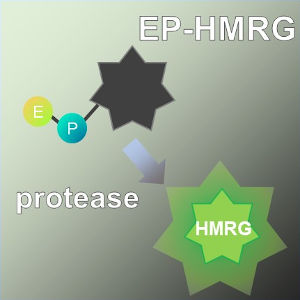
ProteoGREEN™-gGlu
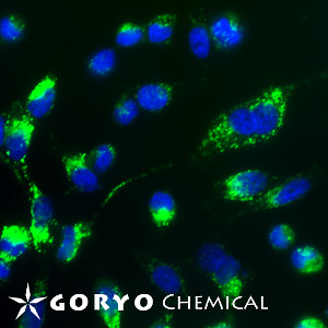
A cell imaging example using ProteoGREEN-gGlu
A cell imaging example using ProteoGREEN-gGlu
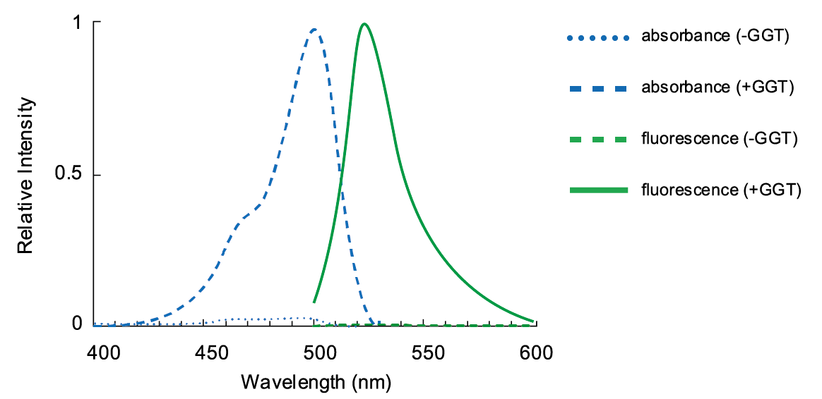
GGT activity detection of HeLa cells
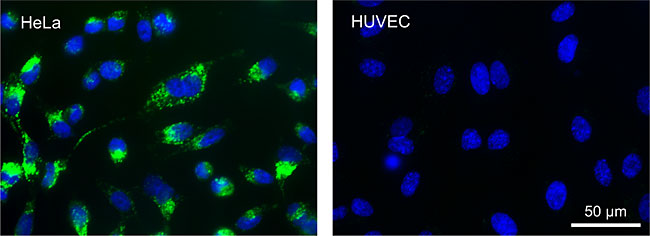
These images are taken 15 minutes after Hela (cell line from human uterocervical cancer, left) cells and HUVEC (human umbilical vein endothelial cell, right) cells were treated with 1 µM ProteoGREEN-gGlu in PBS at 37°C. Green color represents the fluorescence of ProteoGREEN-gGlu and the blue represents the fluorescence of Hoechst 33342, a nuclei marker.
Note that the probe also stains cells without GGT activity, because ProteoGREEN-gGlu-derived fluorophore generated on the GGT-positive cell surface diffuses through the medium and can stains nearby GGT-negative cells. Therefore, this probe is not suitable for distinguishing each cell properties in the mixture of GGT-positive and negative cells.
List of cell lines which application of ProteoGREEN-gGlu has been reported
Cells with enhanced GGT-activity
- Cell lines from human ovarian cancer: SHIN-3, SK-OV-3, OVCAR-3, OVCAR-4, OVCAR-5, OVCAR-8, A2780, A2780 PTX22, IGR-OV1, Hey-A8, CaOV3
- Cell line from human lung cancer: A549
- Cell line from human biliary cancer: HuCCT1
- Cell line from human hepatic cancer: HepG2
- Cell line from human uterocervical cancer: HeLa
- Human breast cancer tissues
Cells with little increase in GGT activity
- HUVEC (Human Umbilical Vein Endothelial Cells)
- SW782 (a non-cancerous cell line of human pre-adipocytes)
- HEK293 cells (Human embryonic kidney 293 cells)

 Powered by Bioz
 Contact Us
