ROSFluor™ Series

HYDROP™ , HYDROP-EX™

[For the specific detection of hydeogen peroxide]

495-540 nm:Green

HYDROP is a fluorescent probe for specific detection of hydrogen peroxide (H2O2). It has low reactivity with other reactive oxygen species (ROS). HYDROP is used for live cell imaging. HYDROP-EX is for detection and quantification of hydrogen peroxide in solutions.


Available through Merck KGaA (Darmstadt, Germany) as:
SCT039 BioTracker™ Green H2O2 Dye  (HYDROP)
SCT040 BioTracker™ Green Free H2O2 Dye  (HYDROP-EX)

Products

Code No. Product Name Size Merck CAT No. Merck ( Millipore / Sigma Aldrich )
Product Name
GC3007-01 HYDROP 30 nmol × 3 SCT039 BioTracker Green H2O2 Dye
GC3008-01 HYDROP-EX 30 nmol × 3 SCT040 BioTracker Green Free H2O2 Dye

Downloads

  • Protocol

  • GC3007 SDS

  • GC3008 SDS

  • Product Information

    Print

    Properties of HYDROP, HYDROP-EX

    Product Name Target Cell permeability Reacivity  Absmax (nm)  FLmax (nm) Photo
    stability
    Brightness
    HYDROP H2O2 Yes (DA) Irreversible 492 516 Low High
    HYDROP-EX H2O2 No Irreversible 492 516 Low High


    • It fluoresces upon reaction with H2O2, but does not react with other ROS such as hydroxyradical (OH), superoxide (O2-・), hypochlorous acid (HOCl), singlet oxygen (1O2), and nitric oxide (NO).
    • Cell-permeable HYDROP is a diacetylated form of HYDROP-EX. Initially, HYDROP has low reactivity with H2O2 but it is quickly hydrolyzed by intracellular esterases to generate reactive and cell impermeable HYDROP-EX. Thus generated HYDROP-EX retains within a cell and fluoresces upon reaction with H2O2.


  • Reactivity of HYDROP-EX and HYDROP

    Print

    Reactivity of HYDROP-EX with reactive oxygen species

    HYDROP-EX shows high specificity to hydrogen peroxide (H2O2).  The reactivity of HYDROP-EX is equivalent to that of HYDROP in cells, because cell-permeable HYDROP is hydrolyzed within cells to generate HYDROP-EX.


    Reaction of HYDROP-EX with various reactive oxygen species. Only H2O2 increases the fluorescence of HYDROP-EX at physiological pH of 7.4 or higher. Fluorescence intensity of 10 µM HYDROP-EX was measured after addition of each ROS (final conc. 50 µM) in 0.1 M sodium phosphate buffer at pH 7.4 containing 0.1 % DMF as a cosolvent. Fluorescence intensities were measured at 520 nm, with excitation wavelength of 490 nm. (Data was kindly provided by Prof. Dr. Y. Urano, Univ. Tokyo)

    ROS generating system

    O2: KO2 ; H2O2: H2O2, 37℃, 60 min ; OH: Fe(ClO4)2:H2O2 =10:1, 37℃, 60 min ; ONOO: HOONO, 25℃, 5 min ; ClO: NaOCl, 25℃, 5 min ; TBHP: tert-Butyl hydroperoxide ; NO: NOC13, 37℃, 30 min ; 1O2: EP-1, 37℃, 30 min

    Quick reaction with H2O2.

    Fluorescence increase can be observed just after the addition of H2O2. Fluorescence intensity increases as the incubation time become longer.

    10 μM of HYDROP-EX was dissolved to 0.1 M phosphate buffer (pH=7.4, including 0.1 % DMF as a cosolvent). H2O2 was added to each concentrations at the timepoint of 5 min (yellow arrow). Fluorescence intensity was measured using a microplate reader with excitation wavelength of 460 nm and emission wavelength of 520 nm at 37℃. Slit widths of excitation and emission was 9 nm and 20 nm, respectively.


  • Live-cell imaging of intracellular hydrogen peroxide using HYDROP™

    Print

    Live-cell imaging of intracellular hydrogen peroxide using HYDROP™

    Imaging of H2O2 production in RAW 246.7 cells

    1. Seeded RAW264.7 cells (5 × 10⁴ cells/mL) on glass bottom dishes and cultured the cells in DMEM without FBS at 37°C in 5% CO2  for overnight.
    2. Diluted the 1 mM HYDROP DMSO solution 1000 times with DMEM without FBS to prepare 1 µM reacting solution.
    3. Added each of the ROS inhibitors (antioxidant) to the reacting solution (Apo; final 5 mM of apocynin , Ebs; final 5 µM of ebselen, NAC; final 10 mM of N-acetyl-L-cysteine).
    4. Removed the culture media and rinsed cells once with HBSS.
    5. Added the reacting solution and incubated for 20 minutes at 37°C, 5% CO2.
    6. Induced hydrogen peroxide production by the addition of final 1 ng/mL of phorbol myristate acetate (PMA) in DMEM without FBS, and incubated for 30 minutes at 37°C, 5% CO2.
    7. Removed the PMA solution from the dish and rinsed cells twice with HBSS. Observed cells with fluorescence microscopy.

    ※ Since the best concentrations of the reagents and the reaction time could be varied depending on the cell type and culture conditions, we performed preliminary tests to choose an appropriate condition. Optimization of the PMA concentration and incubation time is recommended.

    In RAW264.7 cells stimulated with PMA, more spot-like signals of HYDROP was observed compared to intact cells (top). Addition of antioxidants inhibits H2O2 productions (bottom). Bar, 25 μm.

    Microscopy condition: Leica DMI 6000 CS, objective lens: 20×, filter cube: L3 (ex. 450-490, em. 527-530 BP)


    Detection of H2O2 production from autophagy-induced HeLa cells

    1. Seeded HeLa cell (5 × 10⁴ cell/mL) on glass bottom dishs with DMEM containing 8% FBS, and incubated overnight at 37°C, 5% CO2.
    2. Removed the culture media and rinsed cells once with HBSS.
    3. Add DMEM containing 8% FBS to control cells, or Replaced the medium with EBSS without serum, and cultured for 2 hours at 37°C, 5% CO2 to induce autophagy. For control cells, DMEM containing 8% FBS was used.
    4. For the addition of antioxidant, pre-incubate cells with DMEM containing 8% FBS and 10 mM of N-Acetyl-L-cysteine (NAC) for 10 minutes at 37°C, 5% CO2. Concentration of NAC should be kept in the following steps.
    5. Diluted the 1 mM HYDROP DMSO solution to 5 µM, using EBSS or DMEM containing 8% FBS, to prepare reacting solutions.
    6. Removed the media and rinsed cells twice with HBSS and added the reacting solutions. Incubated for 30 minutes at 37°C, 5% CO2.
    7. Rinse the cells twice with HBSS, and observed by fluorescence microscopy.

    HeLa cells in starved conditions induces autophagy and produces H2O2 (Scherz-Shouval, 2007 EMBO J. 26:1749-1760). Compared with control cells (top, right), H2O2 production was enhanced in cells cultured in Earle’s balanced salt solution (EBSS, starved condition, top left). H2O2 was quenched by the addition of antioxidant (lower panels). Scale bar, 50 µm. Cells were observed using Leica DMI 6000 CS, objective lens: 20×, filter cube: L3 (ex. 450-490nm, em. 527-530nm BP).


    Detecting H2O2 production in A431 cell

    1. Seeded A431 cells on glass bottom dishes and cultured in DMEM without serum at 37°C, 5% CO2 until the cells reach ~60% of confluent.
    2. Replaced the culture media with DMEM without FBS, and incubated overnight at 37°C, 5% CO2.
    3. Diluted the 1 mM HYDROP DMSO solution with DMEM without FBS to 5 µM (reacting solution). For the addition of antioxidants, either 5 mM of apocynin or 5 µM of ebselen was added.
    4. Removed the media and rinsed cells once with HBSS, added the reacting solutions and incubated for 10 minutes at 37°C, 5% CO2.
    5. Rinsed cells twice with HBSS, then added EGF (500 ng/mL in DMEM without FBS) and cultured for 30 minutes at 37°C, 5% CO2 to induce the production of hydrogen peroxide.
    6. Removed the medium, rinsed twice with HBSS and observed with fluorescence microscopy.

    H2O2 production by A431 cells is enhanced by the stimulation with epidermal growth factor (EGF). Compared with intact cells (top), stronger signal was detected from EGF stimulated cell (second panel). Fluorescent signal was weaker in the conditions in which ROS inhibitor or antioxidant were added (lower panels). Scale bar, 50 µm. Cells were observed using NIKON Ti-E fluorescent microscope with objective lens: 40×, filter cube: GFP-HQ (ex. 457-487, em. 500-545 BP).


  • A timecourse of ferroptosis observed by fluorescence live cell imaging using FerroOrange and ROSFluor series

    Print

    A timecourse of ferroptosis observed by fluorescence live cell imaging using FerroOrange and ROSFluor series

    Ferroptosis is a cell death which depends on intracellular labile Fe2+. Its mechanism is known as a distinct from that of apoptosis or necrosis. Excess amount of intracellular labile Fe2+ induces generation of reactive oxigen species (ROS) and lipid peroxidation, those lead to cell death. It has also been revealed that ferroptosis is induced in some neurodegerative diseases, and some tumor cells are ferroptosis resistant.

    Here, we tried to detect labile Fe2+ that induces ferroptosis, as well as ROS, by using activatable fluorescent probes of FerroOrange, APF, OxiORANGEand HYDROP, in the timecourse of ferroptosis.

    Visualization of Fe2+ and ROS in the process of ferroptosis

    Erastin was applied to HT-1080 cells to induce ferroptosis, and intracellular labile Fe2+ and ROS were imaged after 3, 6, 9 hours after the application, using the fluorescent probes. FerroOrange (1 μM),  APF (5 μM), HYDROP (1 μM), and OxiORANGE (1 μM) were applied  30 minutes before each of the observation timepoint. Fluorescence signal of FerroOrange which indicates labile Fe2+ was maximum at 3 hours after the induction, whereas fluorescence of other ROS probes: APF which detects hydroxyradical (·OH), hypochlorous acid (HClO), and peroxinitrite (ONOO), OxiORANGE which detects hydroxyradical (·OH), hypochlorous acid (HClO), and HYDROP which specifically detects hydrogen peroxide H2O2 were maximum at 6 hours after the induction. These results indicate that in ferroptosis, ROS increase after the increase in labile Fe2+.

    Fluorescence intensity changes of FerroOrange and APF in ferroptosis-induced HT-1080 cells

    Fluorescence intensity of FerroOrange and APF in erastin-applied HT-1080 cells (upper and middle rows) were shown. Images were overlayed as pseudocolor images (bottom). Magenta indicates fluorescence of FerroOrange, green indicates that of APF. Bar indicates 100 µm.

    Fluorescence intensity changes of OxiORANGE and HYDROP in ferroptosis-induced HT-1080 cells

    Fluorescence intensity of OxiOrange and HYDROP in erastin-applied HT-1080 cells (upper and middle rows) were shown. Images were overlayed as pseudocolor images (bottom). Magenta indicates fluorescence of OxiOrange, green indicates that of HYDROP. Bar indicates 100 µm.


    Experiment procedure

    1. HT-1080 cells of 2 × 10⁵ were seeded on glass bottom dishes and cultured until the cells attached to the bottom.
    2. Erastin (final concentration of 30 μM) was added to the media and cultured for2.5, 5.5, 8.5 hours at 37℃, 5% CO2.
    3. Each probes were added to the media and cultured for 30 minutes.
    4. The cells were rinsed twice with HBSS and observed by fluorescence microscopy.

    ※In different cell culture conditions, the concentrations of the reagents and incubation time might be varied. In this experiment, preliminary tests were needed to optimize those conditions.
    ※If the cell adhesion to the dishes was weak, we recommend to use poly-L-lysine coated dished.

    Actual timecourse of the experiment. Actually, erastin was added in different timepoints and addition of fluorescent probes and observations were performed simultaneously. Since these probes reacts with each target and fluoresces irreversibly, timecourse of ferroptosis was observed by the above procedure.


FAQ

  • Q Can I use DMSO for dissolving OxiORANGE, HYDROP, APF, HPF?
    A

    We recommend to use DMF for these reagents because DMSO is known to be a quencher of one of the reactive oxygen species (ROS), hydroxy radicals. Even though you are intended to detect aother ROS, the ROS could be generated by the signal of hydroxy radicals and usage of DMSO could also decrease the target ROS. Therefore we recommend to use DMF instead of DMSO.


  • Q Tell me a selection guide of ROSFluor series.
    A
    Product code Product name Exmax (nm) Emmax (nm) OH ONOO- HClO H2O2 O2-・ ROS detection in solutions ROS detection in cells
    GC3004-01 OxiORANGE 553 577 + + + +
    GC3006-01 HySOx 553 574 + + +
    GC3007-01 HYDROP 492 518 + +
    GC3008-01 HYDROP-EX 492 518 + +
    SK3001-01 HPF 490 515 + + + +
    SK3002-01 APF 490 515 + + + + +
    SK3003-01 NiSPY-3 490 515 + + +

    ※HYDROP-EX is suitable for detection of extracellular H2O2 since it has low cell permeability.

Reference

Y. Takenaka, I. Inoue, T. Nakano, M. Ikeda, Y. Kakinuma (2022)
FEBS J. 289: 1650-1667 DOI: 10.1111/febs.16249

N. Kurozumi, T. Tsujioka, M. Ouchida, K. Sakakibara, T. Nakahara, S. Suemori, M. Takeuchi, A. Kitanaka,  M. Shibakura, K. Tohyama (2021)
Cancer Sci. 112: 3302–3313. DOI: 10.1111/cas.14982  

N. C. Luong, Y. Abiko, T. Shibata, K. Uchida, E. Warabi, M. Suzuki, T. Noguchi, A. Matsuzawa, Y. Kumagai (2020)
J. Toxicol. Sci. 45: 349-363 DOI: 10.2131/jts.45.349

K. Marunaka, M. Kobayashi, S. Shu, T. Matsunaga, A. Ikari (2019)
Int. J. Mol. Sci. 20: 3869 DOI: 10.3390/ijms20163869

K. Araki, K. Kawauchi, W. Sugimoto, D. Tsuda, H. Oda, R. Yoshida, K. Ohtani (2019)
Commun Biol2: 3 DOI: 10.1038/s42003-018-0246-9

M. Abo, E. Weerapana (2019)
Antioxid. Redox Signal. 30: 1369-1386 DOI:10.1089/ars.2017.7408

K. Ueno, M. Urai, K. Izawa, Y.o Otani, N. Yanagihara, M. Kataoka, S. Takatsuka, M. Abe,
H. Hasegawa, K. Shimizu, T. Kitamura, J. Kitaura, Y. Miyazaki, Y.Kinjo (2018)
Sci Rep. 8: 17406, DOI: 10.1038/s41598-018-35699-4

K. Tomita, Y. Kuwahara, Y. Takashi, K. Igarashi, T. Nagasawa, H. Nabika, A. Kurimasa, M. Fukumoto, Y. Nishitani, T. Sato (2018)
Tumor Biol. 40:1010428318799250 DOI:10.1177/1010428318799250

T. Yamamoto, H. Nakano, K. Shiomi, K. Wanibuchi, H. Masui, T. Takahashi, Y. Urano, T. Kamata (2018)
Biol. Pharm. Bull. 41: 419-426 DOI:10.1248/bpb.b17-00804

K. Tomita, Y. Kuwahara, Y. Takashi, T. Tsukahara, A. Kurimasa, M. Fukumoto, Y. Nishitani, T. Sato (2017)
Biochem. Biophys. Res. Commun. 490: 330-335 DOI:10.1016/j.bbrc.2017.06.044

J. L. Kolanowski, A. Kaur, E. J. New (2016)
Antioxid. Redox Signal. 24: 713-730 DOI:10.1089/ars.2015.6588

H. Guo, H. Aleyasin, B. C. Dickinson, R. E. Haskew-Layton, R. R. Ratan (2014)
Cell Biosci. 4: 64 DOI:10.1186/2045-3701-4-64

M. Abo, Y. Urano, K. Hanaoka, T. Terai, T. Komatsu, T. Nagano (2011)
J. Am. Chem. Soc. 133: 10629-10637 DOI:10.1021/ja203521e

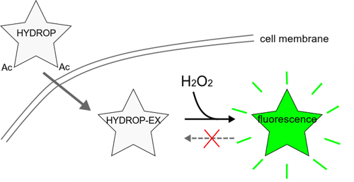
HYDROP is mentioned as NBzFDA.