GlycoFluor™ Series

TokyoGreen®–βGlu

[β-glucosidase activity detection probe]

495-540 nm:Green

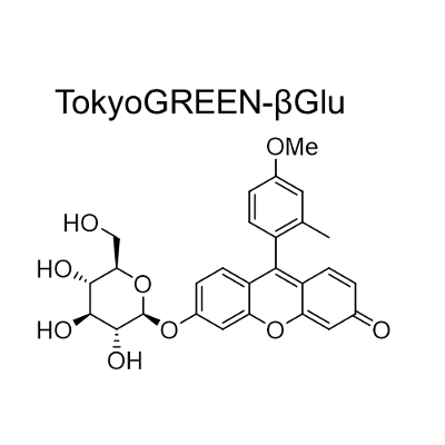
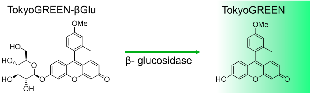
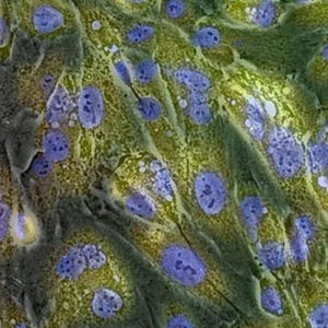
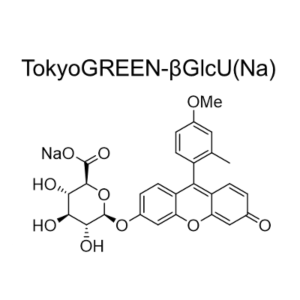
TokyoGreen–βGlu is fluorescent substrate  [9- (4′-methoxy-2′-methylphenyl) -6- (β-D-glucopylanosyloxy) -xanthen-3-one] for β-glucosidase. Almost non-fluorescent TokyoGreen–βGlu is catalyzed by β-glucosidase, and generates bright fluorescent TokyoGreen. It is a cell permeable reagent and can detect intracellular glucosidase. TokyoGreen is also a cell permeable fluorophore, therefore fluorescence is observed both from the cells and extracellular fluid. Thus it is suitable for enzyme assay and for screening using plate reader.


Available through Merck KGaA (Darmstadt, Germany )as:
SCT026 BioTracker™ 510 Green β-Glu Dye

Products

Code No. Product Name Size Merck CAT No. Merck ( Millipore / Sigma Aldrich )
Product Name
SK4002-01 TokyoGreen®–βGlu 1 mg SCT026 BioTracker 510 Green β-Glu Dye
  • Product Information

    Print

    Features

    • Highly sensitive β-glucosidase activity detecting fluorescent probe
    • TokyoGreen, generated by hydrolysis of TokyoGreen–βGlu, has strong fluorescence under the wide range of neutral and basic pH condition.
    • Fluorescent intensity in in proportional to the activity of β-glucosidase.

    Principle of the detection

    Non-fluorescent TokyoGreen–βGlu is hydrolyzed by the β-Glucosidase, and generates bright fluorescent TokyoGreen. TokyoGreen has bright green fluorescence (510 nm) when it is irradiated by the 490 nm excitation light.

    Contents

    TokyoGreen–βGlu 1mg (5 mM in DMSO 0.4mL)
    C27H26O9 Mw:494.49

FAQ

  • Q Give me a selection guide of the β-galactosidase probes.
    A

    We have three β-galactosidase probes, GlycoGREEN-βGal, GlycoGREEN-βGal, and TokyoGreen-βGal . For cell imaging, we recommend GlycoGREEN-βGal, for plate-reader assays or for enzyme kinetics measurements, we recommend TokyoGreen-βGal as a first choice.

    Sensitivity of the probe isGlycoGREEN-βGal ~  TokyoGreen-βGal > GlycoYELLOW-βGal. 
    For cell imaging, we recommend GlycoGREEN-βGal because its cell retention is relatively better, however, generated fluorophore also leaks from the cells and can be incorporated into other cells, users should carefully check the result when they observe heterogeneous cells.  


  • Q Can I use for the fixed samples?
    A

    You may use a similar procedure to X-gal staining for fixed cell samples, because GlycoYELLOW-βGal is also a substrate for β-galactosidase (lacZ reporter)

    We confirmed that fluorescence of GlycoYELLOW-βGal observed in live cells does not diminish by the normal process of fixation. Please refer information in the product page for details.

    Strong fixation conditions may alter the intracellular localization or may decrease the fluorescence. Please test the fixation condition in your cells and conditions.

  • Q My question is not in this FAQ list.....

Reference

Y. Zhou, S. Kajiyama, K. Itoh, T. Tanino, N. Fukuda, T. Tanaka, A. Kondo, K. Fukui (2009)
Appl. Microbiol. Biotechnol. 84: 375-382 DOI:10.1007/s00253-009-2091-8

K. Matsuura, T. Yashiro, K. Shimizu, S. Tatsumi, T. Tamura (2009)
Curr. Biol. 19: 30-36 DOI:10.1016/j.cub.2008.11.030

Y. Urano, M. Kamiya, K. Kanda, T. Ueno, K. Hirose, T. Nagano (2005)
J. Am. Chem. Soc. 127: 4888-4894 DOI:10.1021/ja043919h