NOFluor™ Series

DAR-4M (diaminorhodamine-4M)

[For detection of nitrogen oxide by orange fluorescence]

570-590 nm:Orange

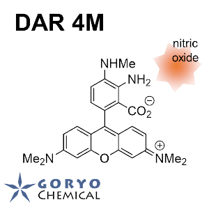
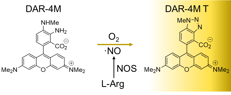
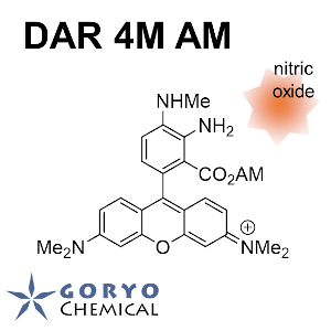
Diaminorhodamine-4M (DAR-4M) is a fluorescent probe to detect nitrogen oxide (NO). This compound alone shows quite weak fluorescence but strongly fluoresces upon the reaction with NO. Compairing with DAF-2 and DAF-FM, it is a improved reagent which fluorescence is less disturbed by autofluorescence of tissues, and it can be used at wider range of pH (4-12). It is cell impermeable and suitable to detect extracellular NO.

Products

Code No. Product Name Size Merck CAT No. Merck ( Millipore / Sigma Aldrich )
Product Name
SK1005-01 DAR-4M 1 mg

Downloads

  • Protocol

  • SDS

  • Product Information

    Print

    Principle of detection

    Nonfluorescent DAR-4M reacts with NO to generate fluorescent DAR-4M T, which can be excited by green light of ~560 nm in wavelength and emits orange fluorescence of ~ 575 in wavelength.

    Properties of DAR-4M

    Product name target reaction pH range Absmax (nm) FLmax (nm) ε Φ
    DAR-4M NO irreversible 4-12 560 575 76,000 0.42

    Spectra

    Product properties

    DAR-4M  1 mg dissolved in 0.47 mL of DMSO.
    C25H26N4O3        
    MW:430.50

    This product is 5 mmol/L solution.
    Dilute ~ 500 times with neutral aqueous buffer solution.


FAQ

  • Q Can I detect the fluorescence with a fixed-wavelength platereader of excitation at 485 nm and detection at 538 nm?
    A

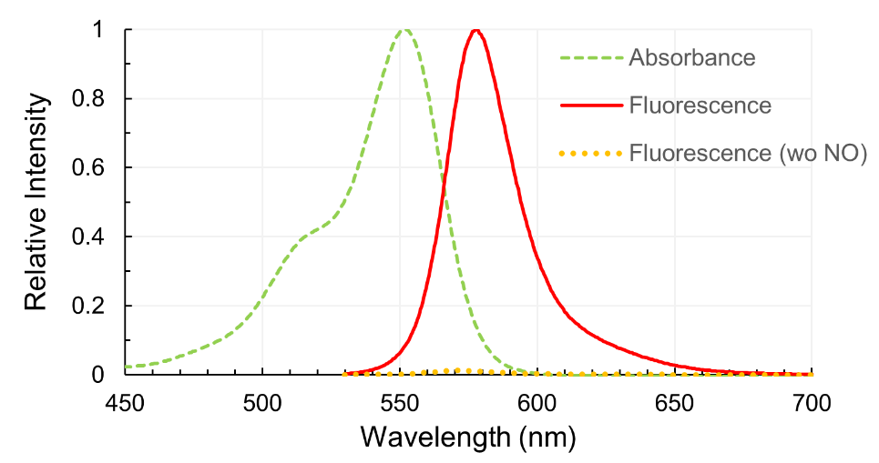
    Yes. Excitation max at 495 nm and fluorescence max at 515 nm are the wavelength of each peaks. It can be measured at the wavelengths of slightly differs (Anal. Chem. 1998, 70, 2450). It is often observed that excitation at the wavelength shorter than the peak and detection at the wavelength longer than the peak lower the background and make better detection efficiency. Also refer the manual of the plate reader since it depends on the properties of the detector.


  • Q What is the detection limit?
    A

    In the buffer of around pH 7 using DAF-2, NO detection limit is 5 nM. In case of using DAF-FM, it becomes slightly high sensitive as its intensity of fluorescence is 1.5 times higher than that of DAF-2. Lower sensitivity can be expected when DAF-2 DA or DAF-FM DA are used to detect intracellular NO because of intracellular  interfering substances.

  • Q Is there any cytotoxicity with these reagents?
    A

    The clear cytotoxicity was not recognized at around the concentration of 10 μM. In case the toxicity might be suspicious, please lower the concentration.

  • Q What is the optimum concentration?
    A

    Optimum concentration is about 5 – 10 μM (diluted by 500 – 1000 times). It could change by the kind of the sample and the buffer used. Due to the properties of the fluorescein-based compound, it may have an adverse effect if the density of the reagent is raised for getting a strong signal.


  • Q NO is a highly reactive compound, but, what state of NO does DAF series detect?
    A

    NO immediately reacts with oxygen, and gives NO2 and NO3. DAFs react with intermediates, which are generated from NO and oxygen.  DAFs can detect NO specifically, because these intermediates cannot be produced without NO generation under physiological conditions.

  • Q Which of DAF/DAR reagents is the most appropriate for my purpose?
    A

    The first choice is DAF-FM for extracellular detection, and DAF-FM DA for intracellular detection.

    DAF-2/DAF-2 DA is often used to reproduce the already reported results, because there are many publications using DAF-2 reagents. In contrast, DAF-FM/DAF-FM DA usually give better results at pH 6-7 range.

    For the detection in a more acidic environment, or when green autofluorescence is not ignorable, DAR-4M/DAR-4M AM are good choices.


    Product code Product name Exmax (nm) Emmax (nm) Cell permeability Capable pH range
    SK1001-01 DAF-2 495 515 >7
    SK1002-01 DAF-2 DA 495 515 + >7
    SK1003-01 DAF-FM 495 515 >5.5
    SK1004-01 DAF-FM DA 495 515 + >5.5
    SK1005-01 DAR-4M 560 575 4-12
    SK1006-01 DAR-4M AM 560 575 + 4-12


  • Q Any possibility to react with nitrite ion and nitrate ion ?
    A

    DAF series will not react with nitrite ion (NO2)and nitrate ion (NO3) under physiological conditions. However, if they are incubated for a long time in the presence of high concentration NO2(≧ 10 mM), slight fluorescence will be observed.

  • Q Let me know example of use for these reagents.
    A

    There are many publications using the reagent. Refer the reference section or publication list. Cultured cell, vascular endothelial cell, cranial nerve system such as hippocampal, peripheral blood mononuclear cell, earthworm ganglion, and plant cell are used for test sample. Image observation with the fluorescent microscope, measuring the fluorescence intensity of the specific points of the image, and measurement with multi-well plate reader measurement.


  • Q Is there any compound interfering the reaction?
    A

    Fluorescent substances such as phenol red and vitamins sometimes interfere the observation. Proteins such as serum and BSA added to medium also sometimes lower the sensitivity of NO detection.

  • Q What is recommended standard compound in case of determination by using calibration curve?
    A

    For the determination of absolute concentration, it is necessary to use the NO gas solution of known density. However, practically, NONOate (NO of 2 molecules is generated by 1 molecule) is recommended to use.

  • Q My question is not in this FAQ list.....

Reference

Araragi, A. Ikeura, T. Uchiumi (2021)
Plant Biotechnol. in press DOI: 10.5511/plantbiotechnology.20.0907a

T. Nagano (2009)
J. Clin. Biochem. Nutr. 45: 111-124 DOI:10.3164/jcbn.R09-66

J. R. Steinert, C. Kopp-Scheinpflug, C. Baker, R. A. Challiss, R. Mistry, M. D. Haustein, S. J. Griffin, H. Tong, B. P. Graham, I. D. Forsythe (2008)
Neuron 60: 642-656 DOI:10.1016/j.neuron.2008.08.025

T. Flores, C. D. Todd, A. Tovar-Mendez, P. K. Dhanoa, N. Correa-Aragunde, M. E. Hoyos, D. M. Brownfield, R. T. Mullen, L. Lamattina, J. C. Polacco (2008)
Plant Physiol. 147: 1936-1946 DOI:10.1104/pp.108.121459

X. Ye, S. S. Rubakhin, J. V. Sweedler (2008)
Analyst 133:423-433 DOI:10.1039/b716174c

E. W. Miller,C. J. Chang (2007)
Curr. Opin. Chem. Biol. 11: 620-625 DOI:10.1016/j.cbpa.2007.09.018

N. N. Tun, C. Santa-Catarina, T. Begum, V. Silveira, W. Handro, E. L. FlohI, G. F. Scherer (2006)
Plant Cell Physiol. 47: 346-354 DOI:10.1093/pcp/pci252

H. Kojima, M. Hirotani, N. Nakatsubo, K. Kikuchi, Y. Urano, T. Higuchi, Y. Hirata, Nagano, T (2001)
Anal. Chem. 73: 1967-1973 DOI:10.1021/ac001136i