POLARIC® Series

POLARIC®-500c6F

[Cell membrane staining probe]

Others

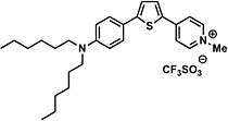
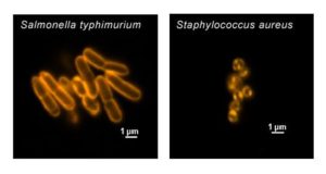
POLARIC-500c6F is solvatochromic dye developed for cell staining. Solvatochromic dye is fluorophore which fluorescence spectrum changes upon polarity of the surrounding solvent.This reagent is modified with alkyl-group which has affinity to cell membranes. Its fluorescence spectrum changes on the membrane lipid component. In most cases, cell membrane and mitochondrial membrane can be distinguished by the difference of fluorescence spectra. 

Products

Code No. Product Name Size Merck CAT No. Merck ( Millipore / Sigma Aldrich )
Product Name
GC101 POLARIC®-500c6F 1 μg × 10
GC1011 POLARIC®-500c6F 1 μg × 5

Downloads

  • Protocol

  • SDS

  • Product Information

    Print

    Properties of POLARIC-500c6F

    Product name target reaction Absmax (nm)* FLmax (nm)*
    POLARIC-500c6F solvent polarity reversible 480 592

    *Because of its low water solubility, it was determined in a aqueous medium containing SDS. It changes upon the solvent polarity.

    Spectra measured in a aqueous medium containing SDS

    POLARIC-500c6F
     

    Fluorescence spectra in cell membrane and mitochondrial membrane

    Spectral data were obtained from confocal laser microscopy of HeLa cells stained with POLARIC-500c6F.


    Structure


    Photostability

    Fluorescence intensities were measured by observing HeLa cells stained with POLARIC-500c6F or MitoTracker Green. Half-life in this observation condition was 70.6 hours for POLARIC-500c6F and 28.6 hours for MitoTracker Green. POLARIC-500c6F enables stable observation for longer time.


    Notes

    POLARIC dyes has been developed by Dr. Koji Yamada (associated professor of Graduate School of Environmental Science, The Hokkaido University), Global Sapporo Bio cluster-S, Regional Innovation Strategy Support Program, Project for Developing Innovation Systems, The Ministry of Education, Culture, Sports, Science & Technology.


  • Cell imaging examples using POLARIC-500c6F

    Print

    Cell imaging examples using POLARIC-500c6F

    HeLa cells stained using POLARIC-500c6F were imaged using confocal microscope with spectral imager. Cell membrane are shown in green pseudocolor and red-shifted mitochondrial membrane shown in orange-pseudocolor is overlayed.


    Mitochondrial membrane imaged by structured illumination microscopy


    HeLa cells stained with POLARIC-500c6F were observed by structured illumination microscope (Nikon, N-SIM).
    Left, conventional fluorescence microscopy; right, 3D-SIM imaging. Ex: 488nm, Em: 510-550nm.

    By using 3D-SIM, which can image ~100 nm structures, detailed structures of mitochondria were observed.


FAQ

  • Q How does POLARIC-500c6F locate in the lipids and fluoresces in response to the lipids?
    A
     

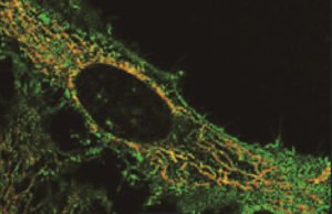
    Because POLARIC-500c6F has a similar structure with lipids, hydrophilic and hydrophobic parts of POLARIC-500c6F orient in pospholipid double membranes. Then POLARIC-500c6F disperses in the membrane to emit strong fluorescence.

  • Q In what situation does POLARIC change the color?
    A

    It is reported that the fluorescent wavelength of POLARIC is changed by the composition of lipid (Chem. Lett. 2011, 4, 989-991). On the other hand, POLARIC show no significant difference of fluorescence wavelength by pH changes.

  • Q My question is not in this FAQ list.....

Reference

M. Takao, Y. Nagai, M. Ito, T. Ohba (2018)
Genes Cells 23: 963-973 DOI:10.1111/gtc.12645

T. N. Mohammadi, A. T. Maung , J. Sato, T. Sonoda, Y. Masuda, K. Honjoh, T. Miyamoto. (2018)
J. Appl. Microbiol. in press DOI: 10.1111/jam.14134

M. Tanaka, T. Kikuchi, H. Uno, K. Okita, T. Kitanishi-Yumura, S. Yumura (2017)
Sci. Rep. 7: 12970 DOI:10.1038/s41598-017-13438-5

R. Omura, K. Nagao, N. Kobayashi, K. Ueda, H. Saito (2014)
J. Lipid Res. 55: 2423-2431 DOI:10.1194/jlr.D049445

N. Maishi, T. Kawamoto, N. Ohga, K. Yamada, K. Akiyama, K. Yamamoto, T. Osawa, Y. Hida, K. Hida (2013)
Oncol. Rep. 30: 1695-1700 DOI:10.3892/or.2013.2620

S. H. Son, Y. Yamagishi, M. Tani, M. Yuasa, K. Yamada (2011)
Chem. Lett. 40: 989-991 DOI:10.1246/cl.2011.989